Jelenlegi hely
Medical Applications
|
The research focuses on investigating the pupillary light reflex to reveal the nervous system changes in rats with schizophrenia-like alterations. |
|
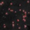
Chlamydiae are obligate intracellular bacteria that propagate in the inclusion, a specific niche inside the host cell. We developed an automatic inclusion counting system based on a commercially available DNA chip scanner. Fluorescently labeled inclusions are detected by the scanner, and the image is processed by ChlamyCount, a custom plugin of the ImageJ software environment. |
|
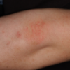
We are researching and developing image processing algorithms for dermatological applications in diagnostic and decision making systems as well as for education. This includes the creation of a personalized surface model from a set of color and depth camera images, detection and classification of dermatological findings, such as psoriasis lesions and plaques, as well as longitudinal analysis of changes. |
|
A novel method for non-rigid registration of transrectal ultrasound and magnetic resonance prostate images based on a non-linear regularized framework. |
|
A novel solution for reassembling a broken object from its parts without established correspondences, where each part is subject to a linear deformation. |
|
We consider the problem of planar shape registration on binary images. Our primary goal is to investigate novel methodologies which work without feature point extraction and established correspondences; avoid the solution of complex optimization problems; and provide an exact solution regardless of the strength of the distortion. The newly developed techniques are validated on medical images. |
|
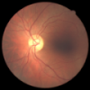
Methods for the automatic detection and evaluation of Glaucoma and other eye diseases from screening examinations. |
|
Our automatic registration method was tailored to the needs of a model-based segmentation framework of the pelvic area in CT studies in collaboration with GE Medical Systems. Registration was used as a preprocesing task. The goal of it was the acceptable alignment of the pubic bone area (where the two organs of interest, the prostate and bladder are located) of studies from different patients. |
|
A joint project is in process between the University of Szeged Image Processing Group and Department of Optics and Quantum Electronics and GE Medical Systems (GEMS) Budapest, to study the problems connected with CT image corrections. |
|
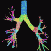
A method for computationally efficient skeletonization of three-dimensional tubular structures was proposeded. It is specifically targeting skeletonization of vascular and airway tree structures in medical images but it is general and applicable to many other skeletonization tasks. |
|
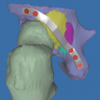
We developed a unified framework for percutaneous therapies utilizing localization frames and implemented an application using CT imaging and a robot. |
|
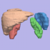
Study and development of image segmentation algorithms for different organs from CT images for radiotherapy planning purposes. |
|
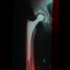
Computer aided surgical planning with semi-automatic segmentation, implant insertion and biomechanical analysis |
|
Skeletonization has been successfully applied in the following three medical applications: assessment of laryngotracheal stenosis, assessment of infrarenal aortic aneurysm, and unravelling the colon. |
|
We developed an image processing method for MRI intensity standardization. We also introduced new, fast implementations of the fuzzy connectedness algorithm that allows segmentation at interactive speeds. We developed a new segmentation "workshop" for brain MRI segmentation using standardized MR images and the fast fuzzy connectedness algorithms. |
|
SZOTE-PACS is the Picture Archiving and Communication System of Albert Szent-Gyorgyi Medical University. It is able to collect studies from CT, MR, SPECT, US and modalities and convert them into DICOM format. |
|
MicroSEGAMS is an AMIGA-based system to perform and evaluate isotope-diagnostic studies. It was created using the experiences with SUPER-SEGAMS. |
|
Supplement to SEGAMS-80 with the creation and evaluation of SPECT studies. It became possible to create user programs for the system in FORTRAN. |
|
The improvement of the SEGAMS system that made possible to automate the image acquisition and evaluation. The system could be generated in different languages as well. |
|
SEGAMS: A tree-structured hierarchical data processing system. The software system of a gamma camera connected to a minicomputer that can be used to acquire and evaluate the most common types of nuclear medicine studies used in clinical routine. |
|
Detecting changes in images acquired by a scintigraph about the distribution of radioactive isotopes is a difficult task. Image processing attempts were made to improve the detection of changes. |

