Jelenlegi hely
Analysis of dermatological images for teledermatology and diagnostic applications

Department of Dermatology and Allergology, University of Szeged
Tandofer Informatika Kft.
We are researching and developing image processing algorithms for dermatological applications in diagnostic and decision making systems as well as for education. This includes the creation of a personalized surface model from a set of color and depth camera images, detection and classification of dermatological findings, such as psoriasis lesions and plaques, as well as longitudinal analysis of changes.
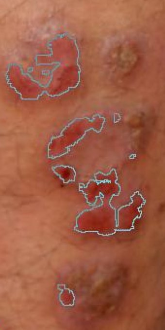
Segmenting psoriasis lesions and plaques
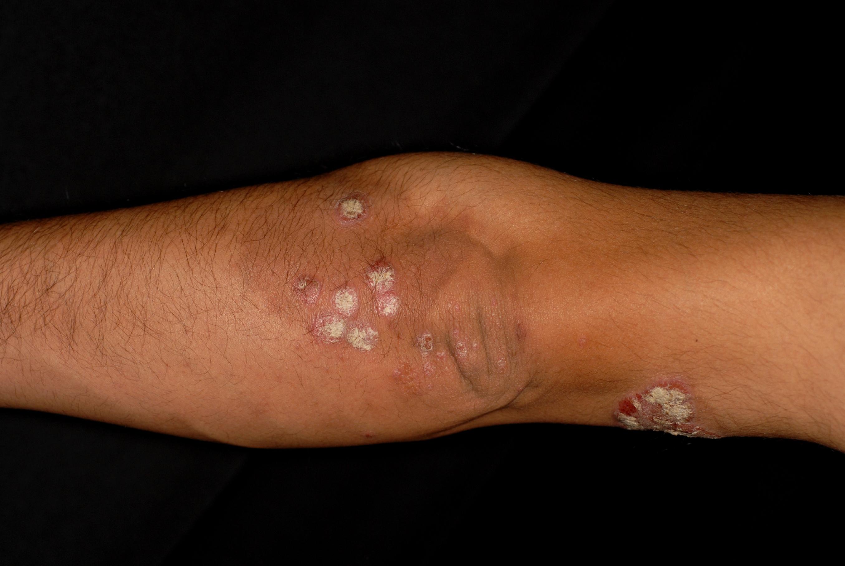
We are studying the possibilities of using the predominant color differences between normal and irritated skin regions as primary features to differentiate. Below are some preliminary examples of segmentation of various types of psoriasis manifestation using different color spaces.
 |
 |
 |
|
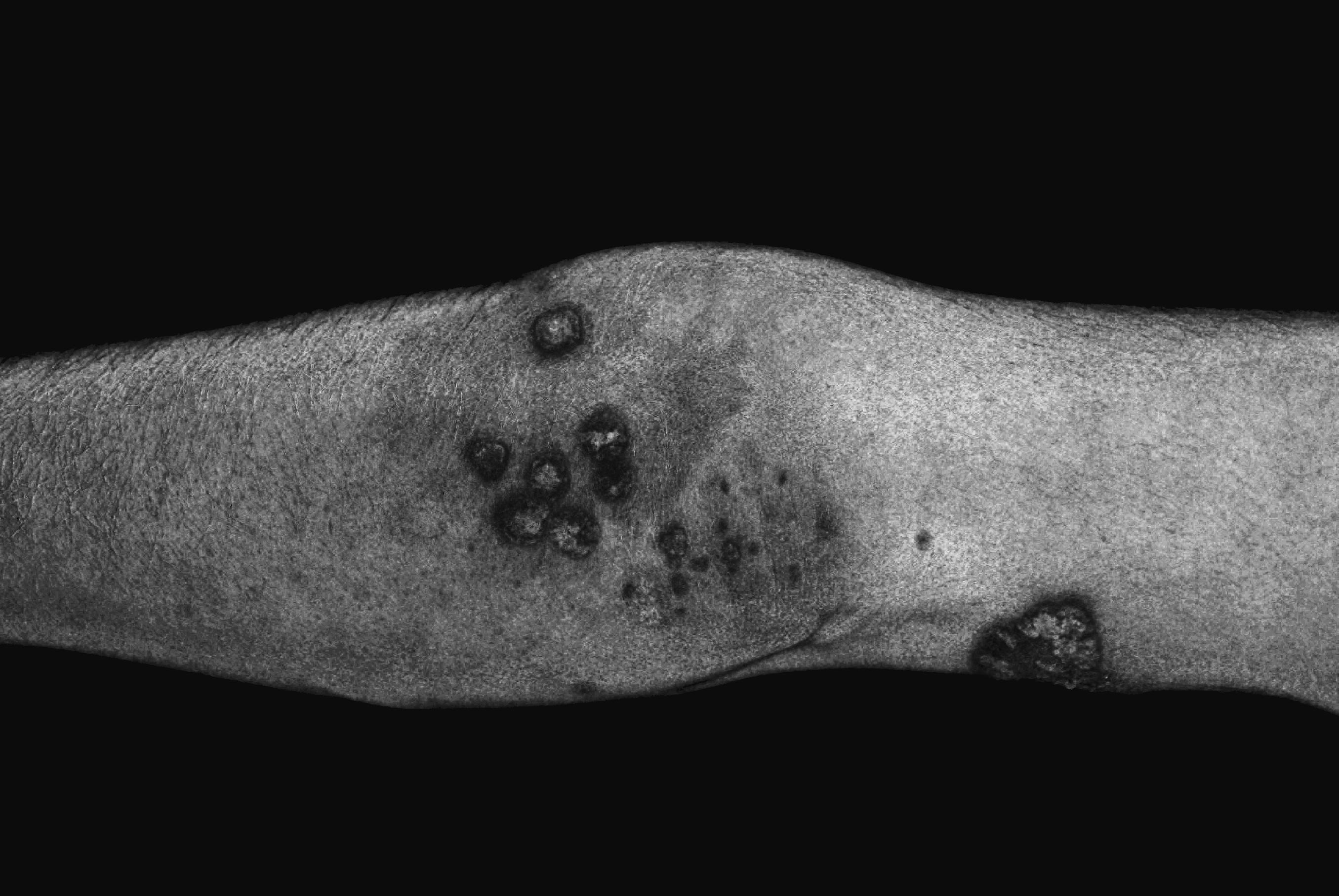
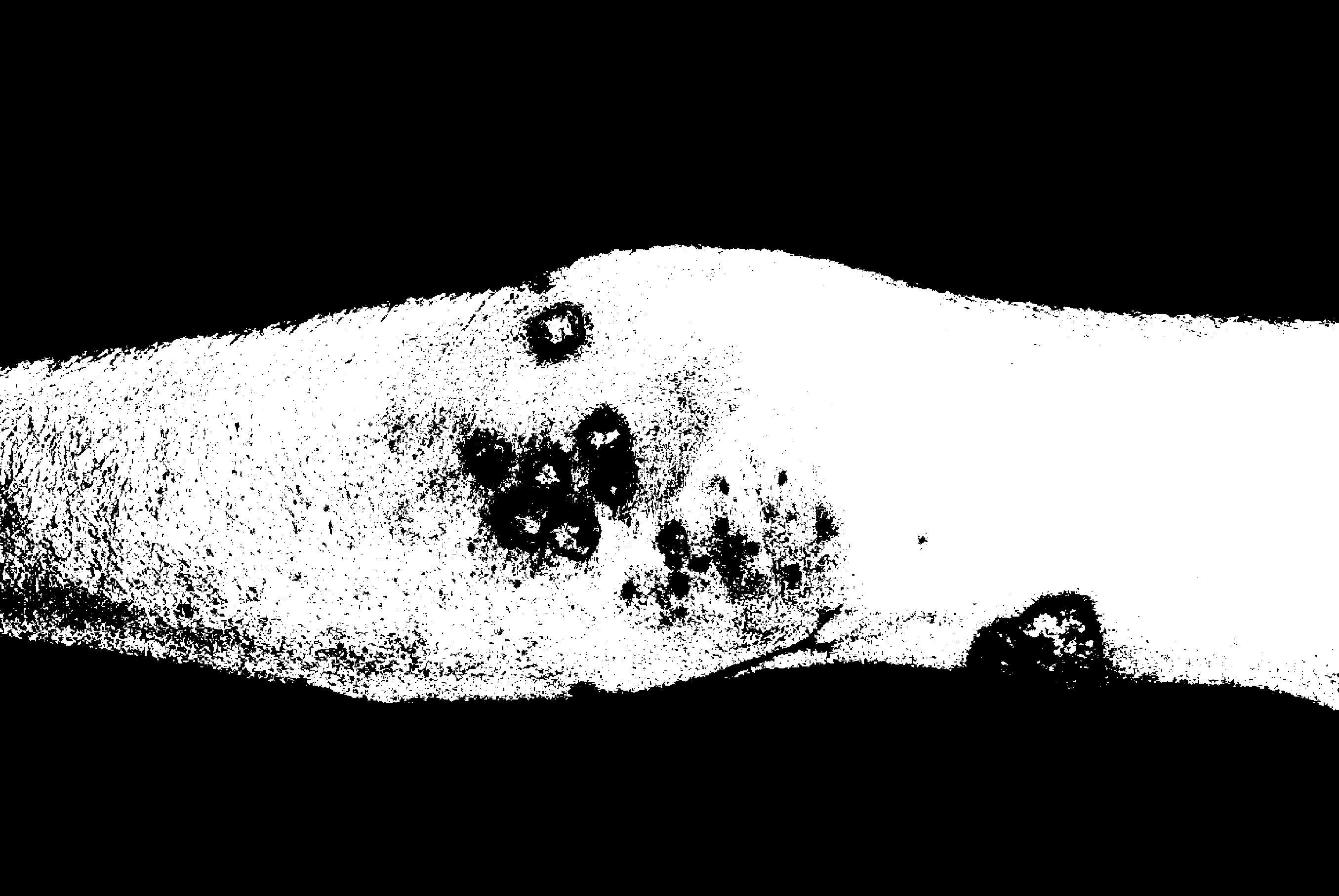
Segmenting irritated regions in the B/G color space. Original image (left), processed color space (center), binary output (right) (without morphological postprocessing).
|
||

Segmenting the plaques (scales) in the Lab color space
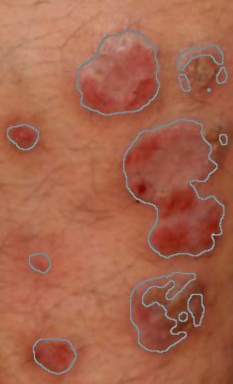
Segmenting irritated red(dish) skin regions in the B-G color space with preprocessing, thresholding, and hole filling
Segmenting irritated red(dish) skin regions in the HSV (H) and YUV (V) color space separately, then thresholding and taking the intersection